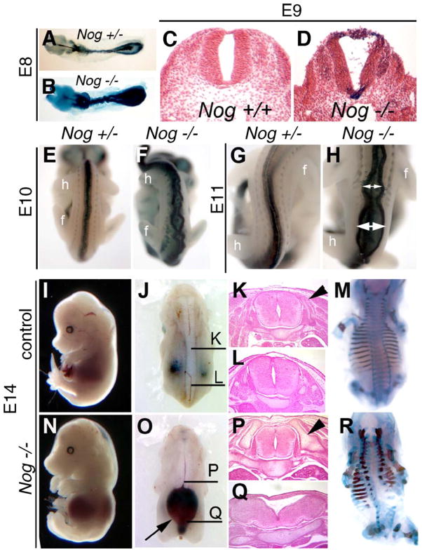
Fig. 4.
Spinal neural tube closure occurs in Nog−/− mutants but is not maintained at trunk levels. (A–D) Whole-mount and histological analysis of control (A and C) and Nog−/− (B and D) embryos at E8 (A and B) and E9 (C and D). E8 mutant embryos show characteristic kinking of spinal cord (B), whereas transverse sections of E9 Nog−/− spinal cords (D) show a distinct neural lumen created by closed neural tube. (E–H) Nog+/− (E and G) and Nog−/− (G and H) embryos at E10 (E and F) and E11 (G and H) show the dorsal neural tube beginning to spread apart at multiple sites in the mutants (double headed white arrows; F and H) in the region between the forelimb (f) and hindlimb (h). (I–R) Wild-type (I–M) and Nog mutant (N–R) embryos at E14. Nog−/− mutants show severe neural tube defects caudal to the forelimb and rostral to the hindlimb, with the dorsal neural tube dramatically separated over an enlarged spinal lumen filled with blood (O, arrow). Histological analysis of control embryos (K and L) shows normal neural tube histology (plane of section indicated in panel J). Nog−/− neural tube at the level of the forelimb looks normal (P) but is dysmorphic more caudally (Q). Dorsal neural arches are forming to surround the neural tube in control embryos (arrowhead in panel K). Nog−/− mutants at the level of the forelimb (P) show enlarged axial skeletal elements (arrowhead in panel P), which do not extend as far dorsally (R). At more caudal levels, the Nog−/− axial skeleton is dysplastic with minimal lateral neural arch formation (Q and R). All paired images are shown at identical magnification.

