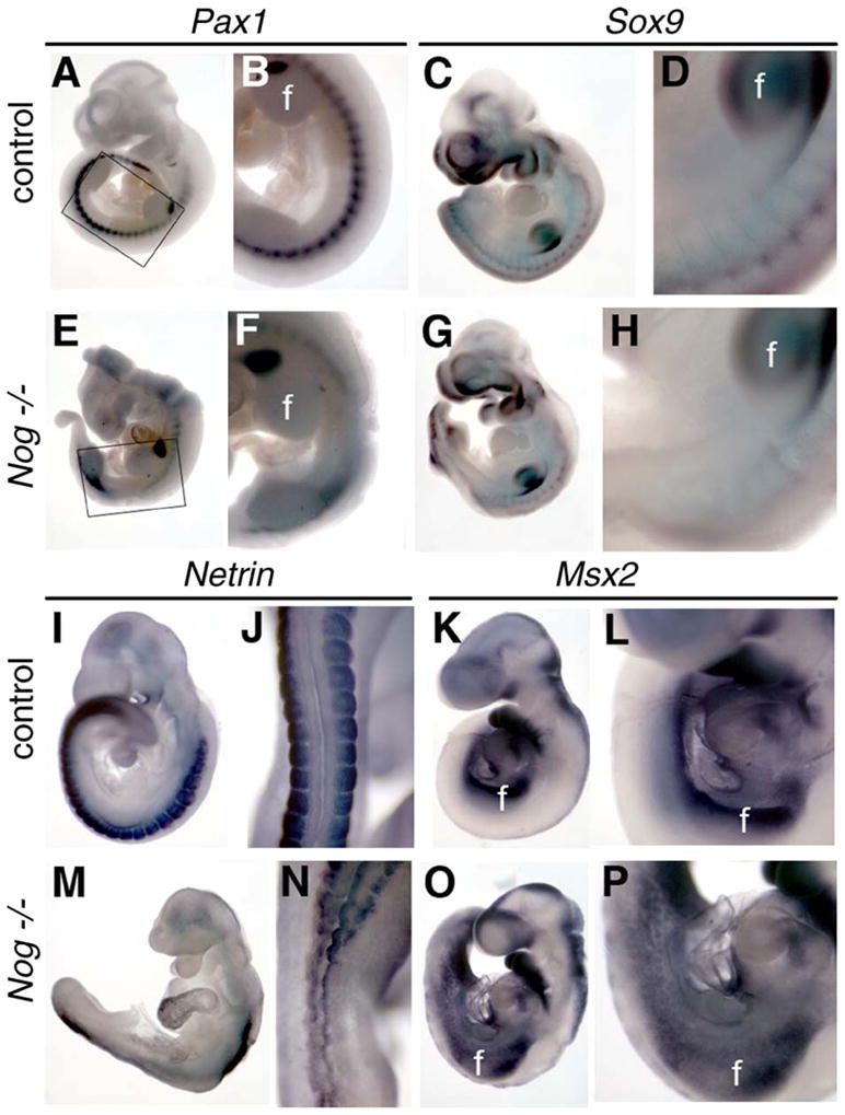
Fig. 5.

Development of the lumbar sclerotome is disrupted in Nog−/− embryos. Whole-mount in situ hybridization for Pax1 (sclerotome marker; A, B, E, and F), Sox9 (marker of cartilaginous differentiation; C, D, G, and H), and Netrin (somitic mesoderm, I, J, M, and N) was used to characterize somitic development in Nog−/− mutants at E9 and E10. Panels J and N show dorsal views, whereas other views are lateral. Pax1 expression in mutants (E, F) was markedly decreased posterior to the forelimb bud (f) and anterior to the hindlimb bud (h). Boxed region in panels A and E is shown at higher magnification in panels B and F, respectively. Similar high magnification views are shown from the forelimb caudally in each pairwise comparison. Sox9 and Netrin expression were also decreased in mutants between the limb buds (G, H, M, and N). (K, L, O, and P) Msx2 expression at E9 is expanded in Nog−/− mutants (O and P) between the limb buds. All paired images are shown at identical magnification.
