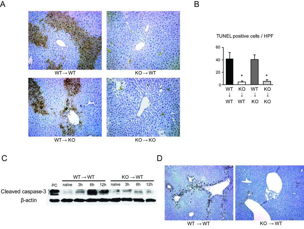
Figure 4. IRF-1 deficiency in liver grafts associated with suppression of hepatocyte apoptosis.
(A) TUNEL staining of liver samples at 12 hours after 4 different LTx. Abundant TUNEL positive hepatocytes (brown) were found in WT→WT and WT→KO LTx. KO→WT and KO→KO LTx showed only few TUNEL positive cells (original magnification ×200).
(B) The numbers of TUNEL positive cells were quantified (n=3 for each group). *p<0.05 vs. WT→WT
(C) Cleaved caspase-3 protein expression in liver grafts. After WT→WT and KO→WT LTx, liver grafts obtained at 3, 6, and 12 hours were analyzed by Western blot. Cleaved caspase-3 expression was significantly less in KO→WT than in WT→WT LTx. Representative images of 3 experiments.
(D) Cleaved caspase-3 staining of liver grafts at 6 hours after reperfusion. Cleaved caspase-3 was expressed on hepatocytes in WT→WT LTx. KO→WT LTx show few positive hepatocytes (original magnification ×100).

