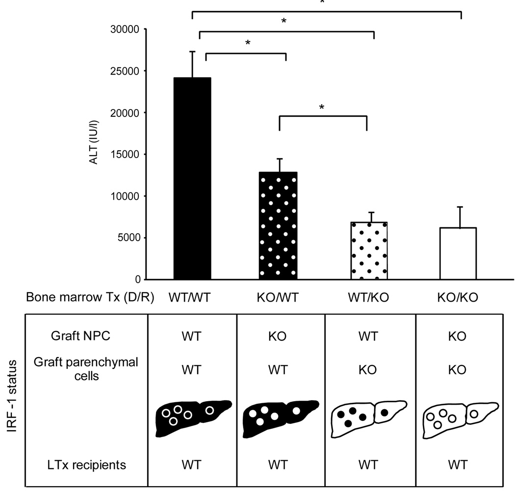
Figure 7. Serum ALT levels of recipients that received liver grafts from bone marrow (BM) chimeras.
Four types of BM chimeras were created in mice. Liver grafts were obtained >2 months later and transplanted into WT recipients, and serum ALT levels of recipients were determined at 12 hours after reperfusion. Liver grafts were from; WT/WT (solid bar): WT BM into irradiated WT mice, KO/WT (black bar with white dots): KO BM into irradiated WT mice, WT/KO (white bar with black dots): WT BM into irradiated KO mice, and KO/KO (white bar): KO BM into irradiated KO mice. IRF-1 status in parenchymal cells (hepatocytes) and NPC was shown for each group. IRF-1 deficiency in NPC (KO/WT) showed significantly lower ALT levels, compared to WT/WT group; however, the lack of IRF-1 in hepatocytes (WT/KO) resulted in further substantial reduction comparable to KO/KO group. (* p<0.05)

