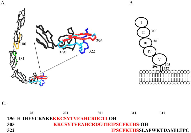
Figure 4. Location of overlapping β2-GPI peptides.
A. Ribbon diagram of human β2-GPI with peptide locations identified by color, peptide 100 (gold), peptide 181 (green), peptide 296 (red), peptide 322 (dark blue) and overlapping peptide 305 (light blue). Inset is magnification of Domain V. B. Cartoon of β2-GPI binding to lipid membrane with peptide locations indicated. C. Sequence identification of overlapping regions of peptides 296, 305 and 322. Red indicates regions of overlap. Peptides were designed based on the published sequences (32) to mimic the lipid binding domain and tail inserted into the lipid bilayer.

