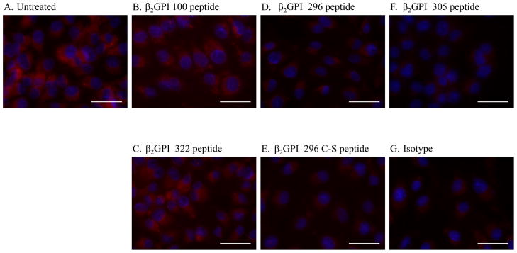
Figure 5. β2-GPI peptides inhibit anti-β2-GPI staining of hypoxic MS-1 cells.
Cells were subjected to 4 hours of hypoxia under serum-free conditions with (A) or without (B–G) β2-GPI peptides prior to 1 hour normoxia in media containing 10% heat-inactivated Rag-1−/− sera. The cells were fixed with methanol, probed with a primary anti-β2-GPI antibody (A–F) or isotype control antibody (G) followed by a Texas red labeled, anti-mouse secondary antibody. Slides were mounted with DAPI (Blue) to identify the nuclei. Each photomicrograph is representative of 3 experiments with 4–6 photomicrographs per treatment in each experiment. Bar = 40μm.

