Figure 3.

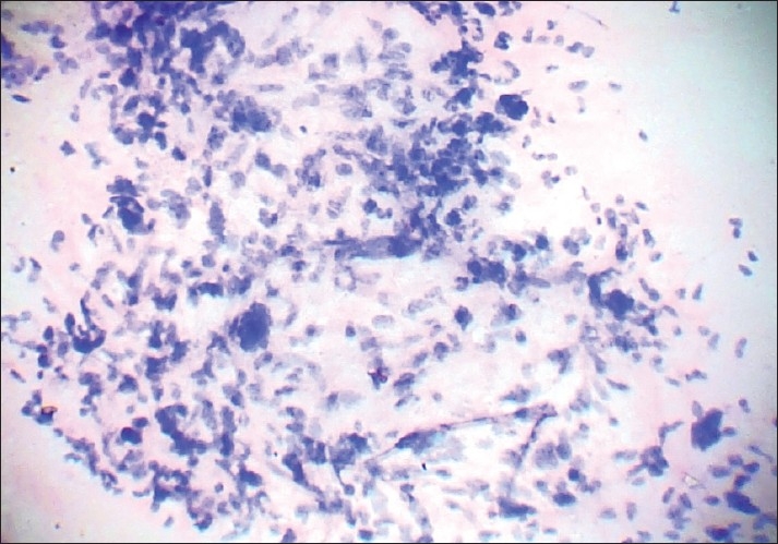
Photomicrograph showing stromal cellular atypia with some bizarre cells in a case of malignant phyllodes tumour. A benign epithelial fragment is visualized in the lower part (PAP, ×400)

Photomicrograph showing stromal cellular atypia with some bizarre cells in a case of malignant phyllodes tumour. A benign epithelial fragment is visualized in the lower part (PAP, ×400)