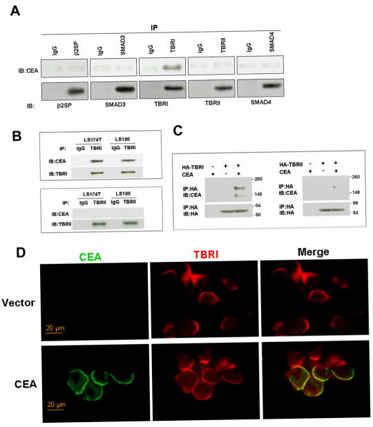
Figure 1. Direct association of CEA with TBRI.
A) 293T cells were co-transfected with wt CEA and one of the five elements of TGF-β signaling pathway, namely β2SP, Smad3, TBRI, TBRII, and Smad4 as indicated. Coimmunoprecipitation experiments were performed to examine the interaction between CEA and these five elements. B) LS174T and LS180 cells lysate were used for the coimmunoprecipitation assay to determine the association of endogenous CEA with endogenous TBRI. C) In vitro binding assay. Purified CEA protein was incubated with purified HA-TBRI or HA-TBRII. Binding of CEA to TBRI or TBRII was assessed by immunoprecipitation followed by immunoblotting. D) 293T Cells were transfected with wt CEA plasmid or vector pcDNA. Cells were then fixed and stained for CEA and TBRI 24 h after transfection.

