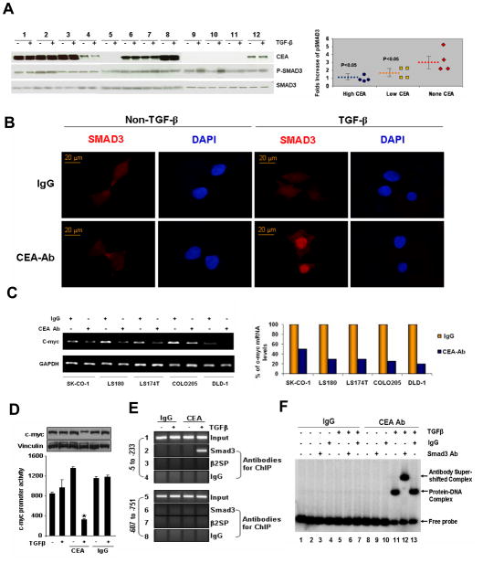
Figure 3. TGF-β signaling is impaired in colorectal cancer cells with elevated CEA.
A) CEA expression and TGF-β induced Smad3 phosphorylation were evaluated in 12 colorectal cancer cell lines by immunoblotting. The extent of Smad3 phosphorylation was measured by the fold increase of p- Smad3 (ratio of p- Smad3 with TGF-β stimulation to p- Smad3 without TGF-β stimulation). According to CEA expression levels, the cell lines were classified into 3 groups. The scatter plot graph demonstrates an inverse correlation between CEA expression levels and the extent of Smad3 phosphorylation. Blue circles: cell lines 1,2,3 and 8; Yellow squares: cell lines 4,6,7, and 12; Red diamonds: cell lines 5,9,10 and 11. Broken lines represent average levels of p-Smad3 fold increase in each group. The bars indicate the standard error. 1. SK-CO-1; 2. LS180; 3. LS174T; 4. Caco-2; 5. HCT-6; 6. Colo205; 7. HT-29; 8. Lovo; 9. HCT116; 10. SW480; 11. HCT-15; 12. DLD-1. B) LS174T cells were treated with anti-CEA antibody (3 μg/ml) or naive IgG for 24 h to block CEA and then treated with or without TGF-β (100 pM) for 1 h. Nuclear translocation of Smad3 was determined as in Figure 2C. C). Five CRC cell lines were treated with or without anti-CEA antibody for 24 h as indicated, then treated with TGF-β (100 pM) for 1 h. Transcription levels of c-myc were assessed as in Figure 2E. The histogram shows the quantification of c-myc mRNA levels in the left graph. D–F) Anti-CEA Ab promotes Smad3-dependent repression of c-Myc expression by TGF-β. D. Effect of IgG or CEA antibody on the c-Myc-luc promoter activity (lower panel) and on the c-Myc protein in the HCT116 cells treated with or without TGF-β. *P<0.05. Western blot analysis was performed with the cell lysates obtained from the luciferase assay samples. E. ChIP analysis showing the recruitment of Smad3 but not β-spectrin onto human c-myc promoter in the HCT116 cells treated with IgG or CEA antibody in the presence or absence of TGF-β treatment. F. EMSA analysis of the Smad3 binding in the human c-Myc promoter using the PCR product encompassing the region −5 to −233 in HCT116 cells treated with TGF-β in presence of either IgG or CEA antibody.

