Figure 2.
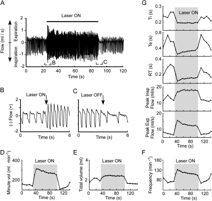
Photostimulation of channelrhodopsin-2–transfected rostral ventrolateral medulla neurons activates breathing: representative plethysmography example. (A) Original trace showing the effect of a 1-minute period of photostimulation (20 Hz, 10-ms pulses, 12 mW) on inspiratory and expiratory flow in a conscious, resting rat. (B) Excerpt from A showing the initial phase of the breathing response. (C) Excerpt from A showing the end of the photostimulation period. Note that the breathing rate and flows were smaller toward the end of the stimulation period than at the beginning. Note also that the frequency and flows were reduced below the prestimulus baseline after interruption of the photostimulation. (D) Change in minute volume caused by the stimulus. (E) Change in tidal volume. (F) Change in breathing frequency. (G) Change in other plethysmography parameters (from top to bottom: inspiratory duration [Ti], expiratory duration [Te], expiratory relaxation time [RT], peak inspiratory flow, and peak expiratory flow).

