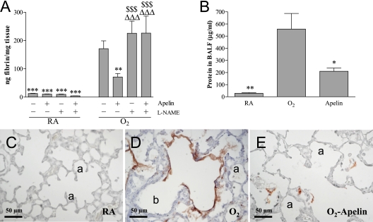
Figure 5.
Quantification of (A) fibrin deposition in lung homogenates, (B) total protein concentration in bronchoalveolar lavage fluid (BALF), and (C–E) expression of β-fibrin on lung sections after early concurrent treatment on Day 10 in room air (RA) and O2-exposed pups (O2) daily injected either with saline, apelin, l-NAME, or a combination of apelin and Nω-nitro-l-arginine methyl ester (l-NAME). Values are expressed as mean ± SEM (n = 10). *P < 0.05, **P < 0.01, ***P < 0.001 versus age-matched O2-exposed control pups. ΔΔΔP < 0.001 versus own RA control pups. $$$P < 0.001 versus apelin-treated O2 pups. a = alveolus; b = bronchus.

