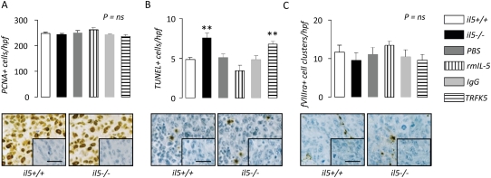
Figure 5.
IL-5 promotes pleural tumor cell survival. Immunodetection of (A) proliferating cell nuclear antigen, (B) TUNEL, and (C) fVIIIra in pleural tumor tissue obtained from wild-type (il5+/+), IL-5 knock-out (il5−/−), and phosphate-buffered saline–, rmIL-5–, IgG-, and TRFK5-treated il5+/+ C57BL/6 mice (n = 7/group) 14 days after intrapleural delivery of LLC cells. Endogenous IL-5 deficiency enhances, exogenous IL-5 delivery decreases, and IL-5 blockade increases pleural tumor cell apoptosis. Microphotographs show representative images of staining from il5+/+ and il5−/− mice (inlays = isotype controls; bars = 50 μm; original magnification ×400; brown = immunoreactivity; blue = nuclear hematoxylin counterstaining). Columns = mean; bars = SE. *P < 0.05, **P < 0.01, and ***P < 0.001, respectively, for comparison with appropriate control. fVIIIra = factor VIII–related antigen; Ig = immunoglobulin; LLC = Lewis lung cancer; PBS = phosphate-buffered saline; PCNA = proliferating cell nuclear antigen; rm = recombinant mouse; TRFK5 = anti–IL-5 neutralizing antibody; TUNEL = terminal deoxynucleotidyl nick-end labeling.

