Figure 6.
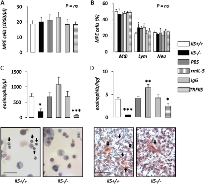
IL-5 promotes eosinophil accumulation in malignant pleural effusion (MPE) and pleural tumors. Total nucleated cells (A), differential cell counts (B), and eosinophil number (C) in MPEs and eosinophil counts in pleural tumor tissue (D) obtained from wild-type (il5+/+), IL-5 knock-out (il5−/−), and phosphate-buffered saline–, rmIL-5–, IgG-, and TRFK5-treated il5+/+ C57BL/6 mice (n = 17, 13, 7, 7, 7, and 11, respectively) 14 days after intrapleural delivery of LLC cells. IL-5 deficiency ablates, exogenous IL-5 enhances, and IL-5 neutralization inhibits eosinophil recruitment to MPE. Microphotographs show representative images of MPE (left, May-Gruenwald-Giemsa stain; bar = 50 μm; original magnification ×400; arrows, eosinophils) and tissue (right, Biebrich scarlet stain; bar = 50 μm; original magnification ×400; arrows, eosinophils) eosinophil staining from il5+/+ and il5−/− mice. Columns = mean; bars = SE. *P < 0.05, **P < 0.01, and ***P < 0.001, respectively, for comparison with appropriate control. Ig = immunoglobulin; LLC = Lewis lung cancer; Lym = lymphocytes; MΦ = macrophages; MPE = malignant pleural effusion; Neu = neutrophils; PBS = phosphate-buffered saline; rm = recombinant mouse; TRFK5 = anti–IL-5 neutralizing antibody.

