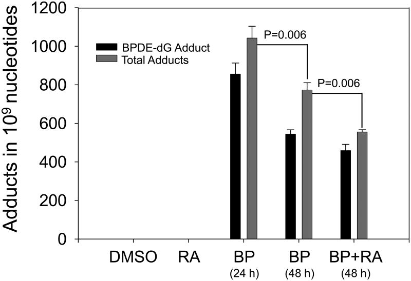
Fig. 4.
Effect of RA treatment on BPDE adduct levels in HepG2 cells. Levels of BPDE-dG and total DNA adducts in cells treated with 4 μM BP for 24 h then incubated another 24 h with DMSO were significantly lower compared to 48 h incubated with 4 μM BP (*P=0.02). After 24 h incubation with 4 μM BaP, the fresh medium was replaced with medium containing 1 μM RA for another 24 h. Total adduct levels were diminished significantly by RA compared to the medium with DMSO in second 24 h incubation (** p=0.032).

