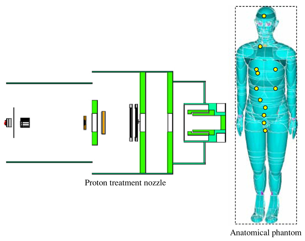
Figure 2.
Schematic illustration of the PSPT treatment unit and the computational anatomical male phantom modeled using Monte Carlo simulations. The locations of the neutron dose receptors are shown as yellow circles on the phantom. The dash line box represents the imaginary box used to enclose the phantom for calculation convenience. The figure is not drawn to scale. (Rendering of anthropomorphic phantom was provided courtesy of Tom Jordan.)

