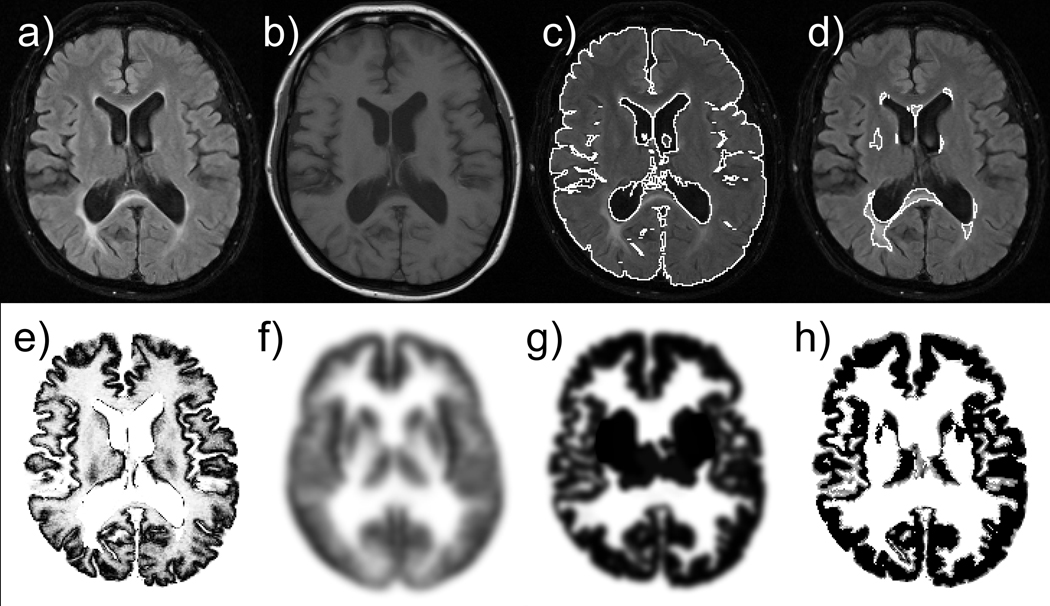
Figure 2.
Example of input (a–b), intermediate (c–g) and final segmentation (h) images: a) input FLAIR image, b) input T1-weighted image, c) brain segmentation, d) lesion segmentation, e) intensity-based probability map, f) anatomy-based probability map, g) morphology-based probability map, and h) resulting GM mask.

