
FIGURE 4. Effect of ERK inhibition on ox-PAPC inhibitory effects.
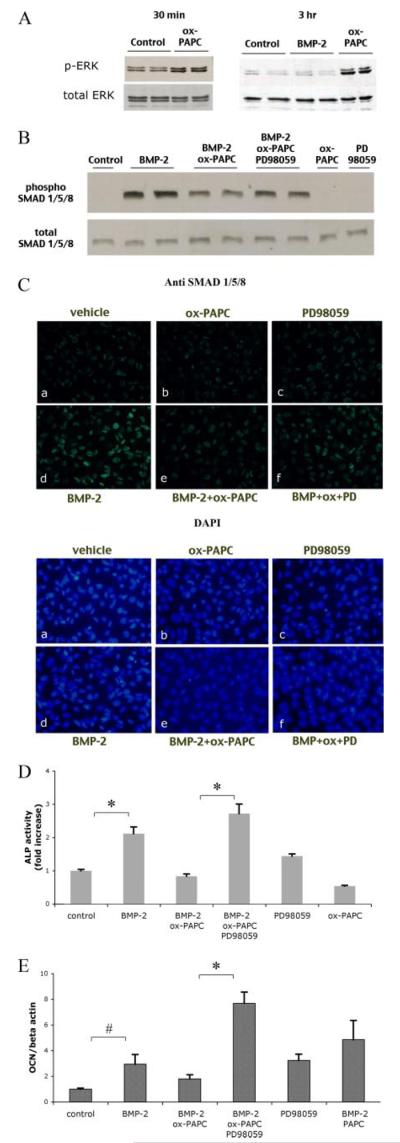
A, activation of ERK in response to BMP-2 and ox-PAPC. Western analysis of phosphorylated ERK (p-ERK) from cell cultures treated for 30 min (left panel) or 3 h(right panel) with BMP-2 (50 ng/ml) or ox-PAPC (10 μg/ml). Total ERK was used as a loading control. B, activation of SMADs in response to BMP-2, ox-PAPC, and PD98059. Western analysis of phosphorylated SMAD 1/5/8 in cells treated for 1.5 h with vehicle, BMP-2 (100 ng/ml), ox-PAPC (30 μg/ml), or PD98059 (5 μm), BMP-2/ox-PAPC, or BMP-2/ox-PAPC/PD98059. Total SMAD 1/5/8 was used as a loading control. C, immunofluorescent (fluorescein isothiocyanate) staining of phosphorylated SMAD 1/5/8 (green; upper panels) in cells pretreated for 30 min with PD98059, followed by treatment for an additional 1.5 h with vehicle (a), ox-PAPC (b), PD98059 (c), BMP-2 (d), BMP-2 and ox-PAPC (e), BMP-2, ox-PAPC, and PD98059 (f) in medium supplemented with 0.5% fetal bovine serum. 4 ,6-Diamidino-2-phenylindole staining (blue; lower panels) was used to visualize cell nuclei (magnification ×400). D, alkaline phosphatase activity in response to BMP-2, ox-PAPC, and PD98059. Cells were treated for 2 days with BMP-2 (50 ng/ml), ox-PAPC (10 μg/ml), and/or PD98059 (5 μm) as indicated. E, osteocalcin expression in response to BMP-2, ox-PAPC, and PD98059. Cells were cotreated for 3 days with vehicle, BMP-2 (50 ng/ml), ox-PAPC (10 μg/ml), PAPC (10 μg/ml), or PD98059 (5 μm) as indicated. *, p < 0.0001; #, p < 0.05.