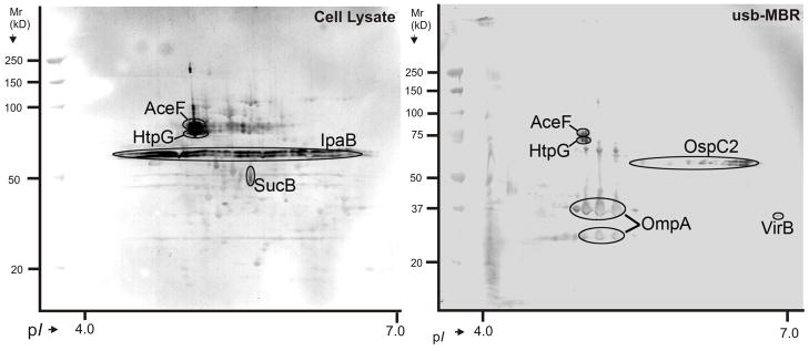
Fig. 6.
S. dysenteriae serotype 1 antigens detected with a piglet antiserum in a 2D western blot. Left: cell lysate; right: usb-MBR fraction. Matching CBB-stained 2D gels were used to identify the spots by MS. Thorough validation was required to designate a protein confidently as an antigen: (1) highest LC-MS/MS score from at least two gel spots matching the western blot signal; (2) same spot shape in 2D gel and western blot; (3) availability of MALDI-TOF data (IpaB and VirB were the exceptions); (4) cross-validation of data via SDS-PAGE western blotting after fractionation of a cell lysate by anion exchange chromatography (data not shown here).

