
Figure 2.
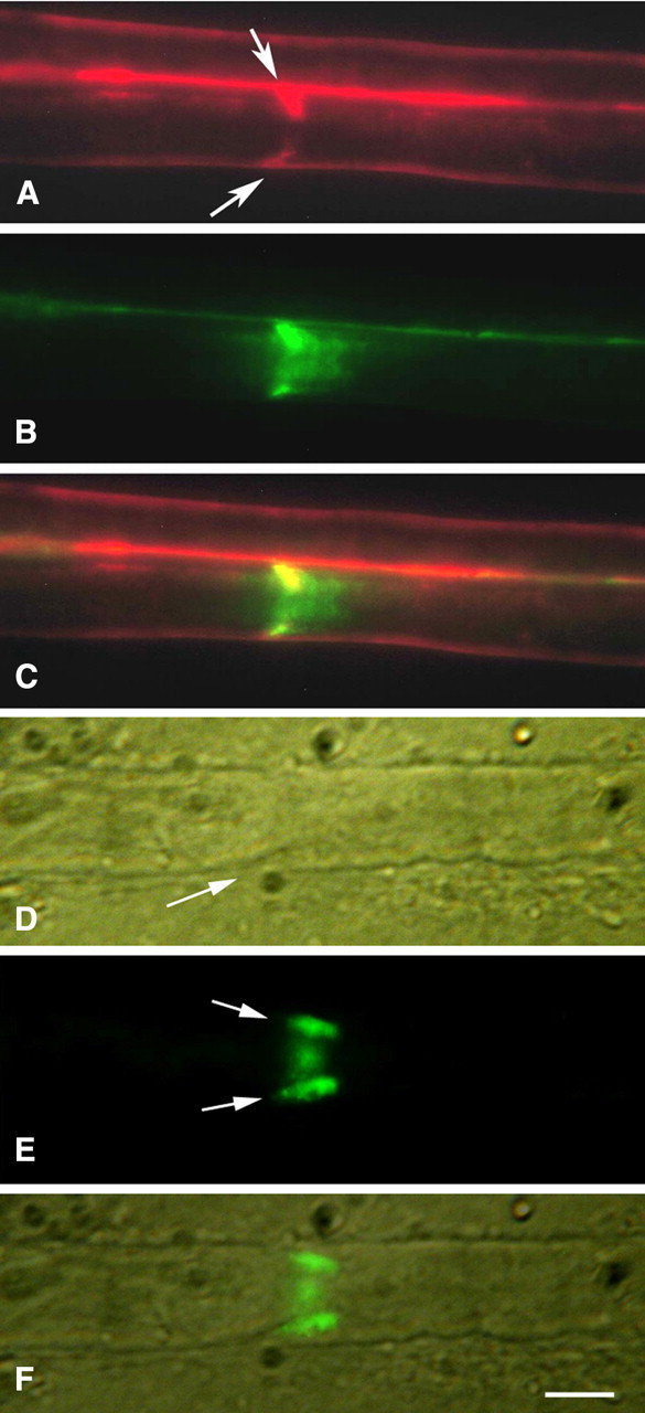
A–C, Schmidt–Lanterman cleft (live injection). Both tracers, 70 kDa (red) and 3 kDa (green), outline the outer surface of the fiber and both extend obliquely from the fiber surface toward the axon (arrows). The 3 kDa tracer then spreads longitudinally along the axon in both directions (B). The 70 kDa tracer (A) does not extend beyond the inner end of the cleft. C, Merged A and B images. In A and C, the red outline of a second fiber is visible above the outline of the first. D–F, Schmidt–Lanterman cleft in fixed fiber exposed to 70 kDa dextran (green) for 4 h. Arrows in E show the tracer extending obliquely into the cleft (D, arrow) toward the axon from both sides of the fiber and visible also in the frontal view between. F, Merged D and E images. Scale bar, 5 μm.