
Figure 8. Summary of residues that exhibit significant conformational exchange for apo and Ca2+-bound C-CaM in the presence of PEP-19.
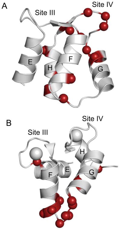
Panels (A) and (B) show the NMR solution structures for apo (pdb: 1F71) and Ca2+-bound C-domain of CaM (pdb: 1J7P), respectively. Colored balls indicate residues that exhibit significant conformational exchange, and are shown by bold lettering in Table 2.