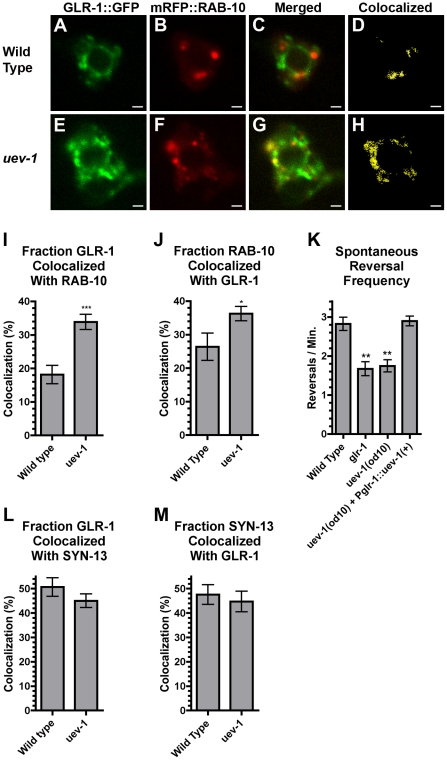
Figure 6. UEV-1 regulates GLR-1 colocalization with endosomal protein RAB-10.
(A, E) GLR-1::GFP and (B, F) mRFP::RAB-10 fluorescence was observed in single-plane confocal images of PVC neuron cell bodies from (A–D) wild-type animals and (E–H) uev-1(od10) mutants. (C, G) Merged images. (D, H) Binary masks (yellow) were created to highlight pixels with matching intensity values for both GLR-1::GFP and mRFP::RAB-10, indicating colocalization. The mean percent of (I) GLR-1 colocalized with RAB-10, and (J) RAB-10 colocalized with GLR-1 is plotted for the indicated genotypes. More GLR-1 is found colocalized with RAB-10 in uev-1(od10) mutants. (K) The mean spontaneous reversal frequency as an indication of GLR-1 function is plotted for the indicated genotypes. The mean percent of (L) GLR-1::GFP colocalized with Syntaxin-13::mRFP, and (M) Syntaxin-13::mRFP colocalized with GLR-1::GFP is plotted for the indicated genotypes. Bar, 1 microns. Error bars are SEM. N = 20–30 animals for each genotype. (I, J) ***P<0.0001, *P<0.05 by t-test. (K) **P<0.01, *P<0.05 by ANOVA with Dunnett's multiple comparison test to wild type.

