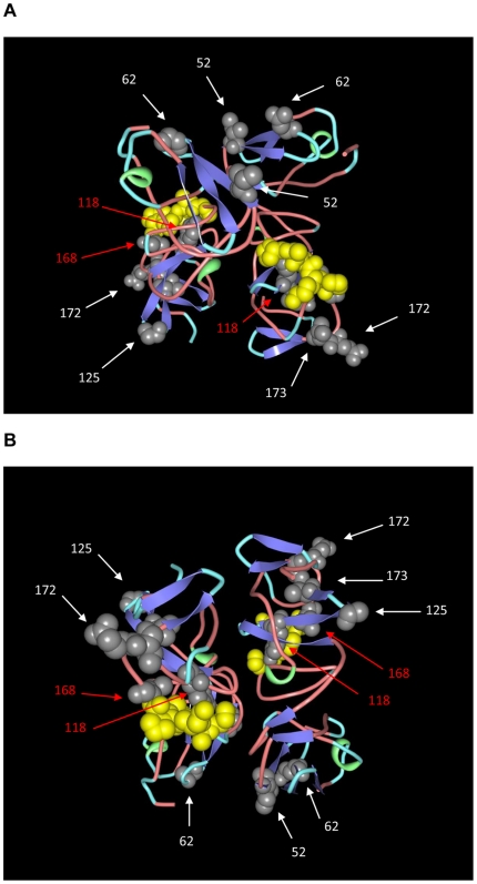
Figure 7. 3D model of domain I of NS5A protein.
A dimeric model of domain I NS5A protein (PDB accession number 3FQQ) is shown. The molecules are colored according to conformational type (turn is shown in light blue, coil in light red, helix in green and strand in blue). Amino acid positions corresponding to NS4A interactions are shown in space filling representation in yellow. Amino acid substitution positions found in C1292 (G2j) are shown in space filling representation in grey and their positions are indicated by numbers and shown by arrows. Red arrows indicate residues spatially close to those known to interact with NS4A protein. Two views of the molecules, rotated under the x-axis are shown in (A) and (B), respectively.

