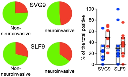
Figure 7. Cytolytic responses of CD8+ cells stimulated with the SGV9 immunodominant WNV peptide are augmented in subject with neuroinvasive involvement.
PBMC from donors with or without neuroinvasive disease were stimulated with the immunodominant SVG9 or subdominant SLF9 peptide WNV epitopes and functional responses were evaluated as described. Percentages of CD8+ T cells expressing the cytotoxicity surrogate marker CD107a were added for each of the 32 combinations shown in Figure 5 using SPICE software. Pies represent the distribution of CD107a positive (red) and negative (green) for the two epitopes. Bars represent the interquartile range (25% –75%) of the CD107a response for the two cohorts. Wilcoxon signed-rank test was performed for the two antigens.

