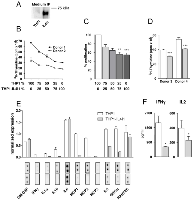
Figure 1. IL4I1 expression by monocytes inhibits T cell proliferation and inflammatory cytokine and chemokine secretion.

(A) Immunoprecipitation of IL4I1 protein from medium of THP1 and THP1-IL4I1 transfectant cells. (B, C, D) Human PBMC from two different donors co-cultured with irradiated THP1 or THP1-IL4I1 cells were stimulated with an anti-CD3 antibody (B & C) or with PPD (D). Proliferation was measured by 3H-thymidine incorporation during the last 18 hours of a 4-day culture. Results in B are expressed as the average cpm of quadruplicates ± standard deviation (SD) after background proliferation subtraction (see methods) of a representative experiment. Panel C depicts percent proliferation of PBMC cultured with THP1-IL4I1 as compared to PBMC cultured with THP1 cells (mean ± SD of independent experiments). (E) PBMC were co-cultured as in B and culture media harvested at day 3 were analyzed on a Raybiotech cytokine array. (F) PBMC were co-cultured as in B and day 1 and day 3 culture media analyzed respectively for IL2 and IFNγ secretion by ELISA (mean ± SD of 7 and 4 independent experiments, respectively). In D, E and F, white bars and gray bars represent results obtained with THP1 and THP1-IL4I1 cells, respectively. * p < 0.05, ** p < 0.01, *** p < 0.001.
