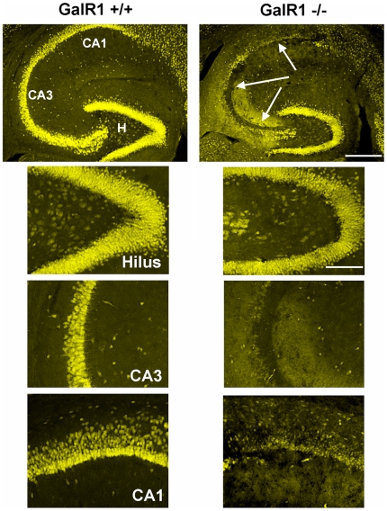
Figure 4. GalR1 deficient mice show increased susceptibility to seizure-induced cell death.
Corresponding low-power and high-power photomicrographs of NeuN-immunofluorescent stained horizontal sections of the hippocampus depicting surviving cells throughout the hippocampus 7 days following systemic KA administration to GalR1−/− and GalR1+/+ mice. Hippocampal sections from GalR1+/+ and GalR1−/− brains were stained with NeuN immunofluorescence to determine the amount of cellular damage. Note the massive loss of neurons, as evidenced by loss of immunostaining, in the hilar, CA3 and CA1 fields of the hippocampus, seven days after a systemic injection of KA inGalR1−/− mice. In contrast, hippocampal cell death was essentially non-existent throughout all hippocampal subfields in GalR1+/+ mice. CA1 and CA3 denote the hippocampal subfields; H, dentate hilus. Scale bars = 750 µm (top panels); 100 µm (bottom panels).

