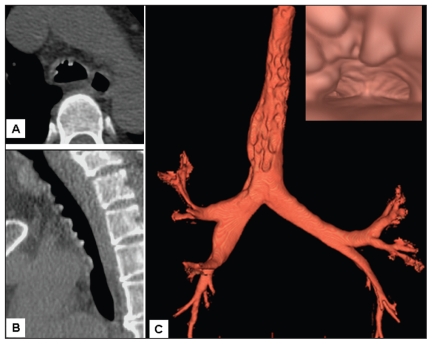
Figure 1:
Chest multidetector computed tomography in a 52-year-old woman with chronic cough. Transaxial section shows multiple calcified nodules at the anterolateral wall of the trachea (A), and sagittal section of the trachea shows typical sparing of the posterior membrane (B). Three-dimensional volume-rendered imaging, functioning as virtual bronchoscopy, shows protruding nodules along the whole trachea (C); the nodules look like stalactites (insert).

