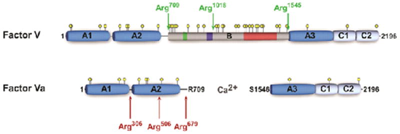
Figure 1. Schematic representation of FV and FVa.

Schematic A1-A2-B-A3-C1-C2 domain representation of human FV and FVa. Thrombin cleavage sites are indicated by green arrows and APC cleavage sites by red arrows. Yellow circles represent potential N-linked glycosylation sites, the green box indicates a 2× 17 amino acid repeat region, the dark blue box corresponds to the basic sequence 963-1008 implicated in preserving the FV procofactor state, and the red box represents a 31× 9 amino acid tandem repeat region.
