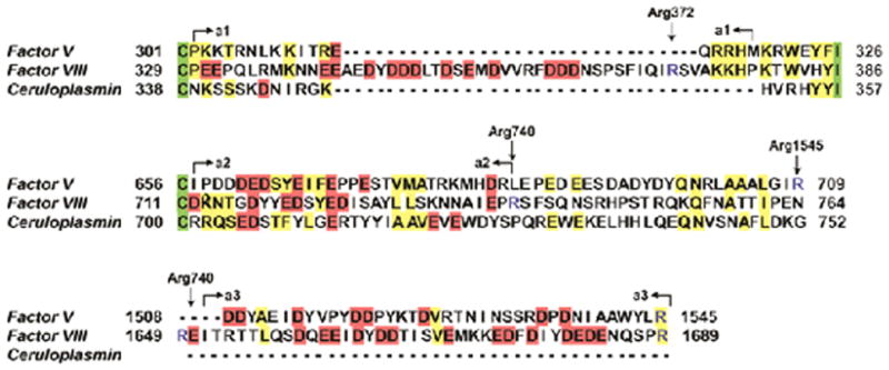
Figure 3. Alignment of the acidic regions a1, a2, and a3 of FVIII.

The acidic regions a1, a2, and a3 from human FVIII were aligned with human FV and human ceruloplasmin (AlignX Module; Invitrogen Carlsbad, CA, USA). Residues fully conserved between all three molecules are shown in green, partially conserved amino acids are indicated in yellow, and the negatively charged residues characterizing the acidic regions are shown in red. The boundaries of the acidic regions are indicated by arrows and thrombin cleavage sites are indicated by arrows and depicted in blue.
