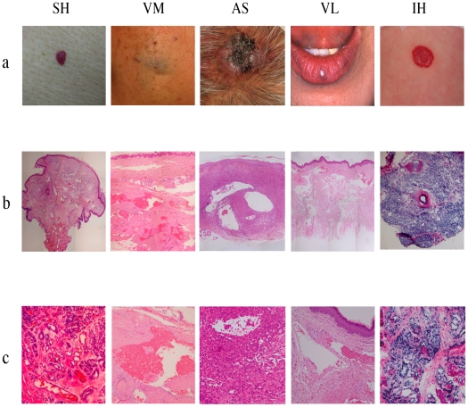
Figure 1. Clinical presentation and histological findings of vascular anomalies.
Panel (a) shows clinical pictures of senile hemangioma (SH), vascular malformation (VM), angiosarcoma (AS), venous lake (VL) and infantile hemangioma (IH). Panel (b) (magnification, ×10) and (c) (magnification, ×100) show hematoxylin-eosin staining of each anomalies. SH showed clusters of proliferated small vascular channels in upper dermis and dilated vessels in the deep dermis. AS consisted of diffuse proliferation of atypical ECs accompanied with irregular vessel-like spaces. Diffuse proliferation of tumor cells and dilated vessels were seen in IH. VM and VL were characterized by thin-walled, dilated vessels accompanied with thrombosis throughout the dermis.

