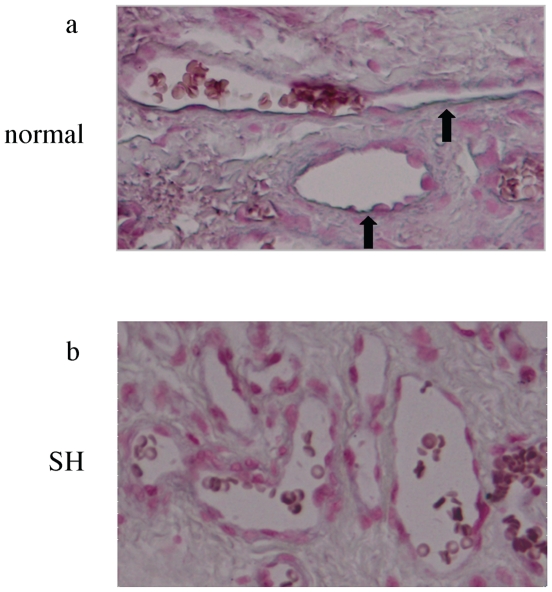
Figure 4. The immunoreactivity for mir-424 in senile hemangioma.
In situ detection of mir-424 in paraffin-embedded, formalin-fixed tissues of normal skin (a) and senile hemangioma (SH, b). Nucleus was counterstained with nuclear fast red (magnification, ×200). The immunoreactivity for mir-424 (blue) is indicated by arrows. Results are representative of 3 normal skins and 3 SH.

