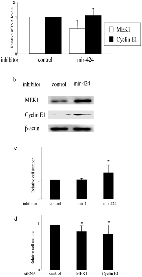
Figure 7. The association of mir-424 with MEK1 or cyclin E1.
(a, b) mir-424 inhibitor induces the protein expression of MEK1 and cyclin E1 but not mRNA expression in human dermal microvascular ECs. Cells were transfected with the control inhibitor or mir-424 inhibitor for 48 hours. (a) Relative amounts of transcripts (normalized with GAPDH) determined in total RNA with quantitative PCR. Error bars represent SD of +1. (b) Lysates from cells transfected with control or mir-424 inhibitor were subjected to immunoblotting with antibody for MEK1 or cyclin E1. The same membrane was then stripped and reprobed with anti β-actin antibody as a loading control. (c, d) mir-424/MEK1/cyclin E1 pathway altered the cell proliferation activity. (c) HDMECs at a density of 5×10³ cells/well in 24-well culture plates were transfected with control inhibitor or inhibitors specific for mir-424 or mir-1. After 48 hours, the number of cells was counted with a Colter® Particle Counter (Beckman Coulter, Fullerton, CA) as described in ‘Materials and Methods’. The value in the cells transfected with control inhibitor were set at 1. The mean and SD from 3 separate experiments are shown. * P<0.05 in comparison to the value in the cells transfected with control inhibitor. (d) HDMECs at a density of 1×10⁴ cells/well in 24-well culture plates were transfected with control siRNA or siRNA specific for MEK1 or cyclin E1. After 72 hours, the cell number was counted as described in Fig. 7c.

