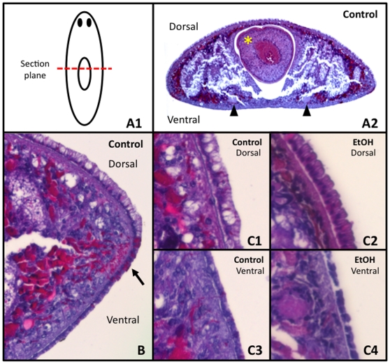
Figure 6. A Short 3% Ethanol Treatment Does Not Cause Epithelial Cell Lysis.
H&E Staining Analysis. (A1) Diagram of sections. (A2) Transverse section of control worm. Yellow asterisk = pharynx. Black arrowheads = round ventral nerve cords (which denote the ventral surface). (B) Closer view of an untreated control section, illustrating the larger columnar dorsal epithelial cells, the smaller cuboidal ventral epithelial cells, and the adhesive cells that lie at the dorsal/ventral margin which stain bright pink (arrow). (C1) Control dorsal, (C2) EtOH-treated dorsal, (C3) control ventral and (C4) EtOH-treated ventral epithelia showing that EtOH treatment does not disrupt epithelial cell membranes.

