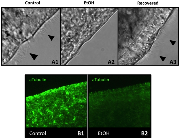
Figure 7. A 3% Ethanol Treatment Removes Epithelial Cila.
(A) Analysis of Cilia in the Head Region. (A1) Untreated control worms are ciliated. (A2) After a 1-hour EtOH treatment, worms have become deciliated. (A3) After 4 hours of recovery, EtOH-immobilized worms are again ciliated. n = 11 for each. Black arrowheads = cilia. (B) Anti-acetylated tubulin staining of cilia on the ventral surface. (B1) Control worms are heavily ciliated (bright green staining). (B2) After a 1-hour EtOH exposure, epidermal ciliary staining is lost. Pre-pharyngeal region is shown.

