Figure 5.
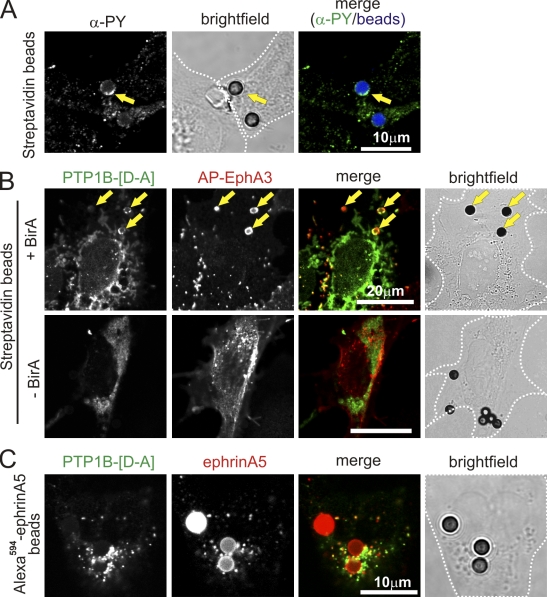
PTP1B interacts with EphA3 at the cell surface. (A) EphA3 clustering by SA beads provokes tyrosine phosphorylation. COS7 cells, cotransfected with APN-EphA3 and BirA, were stimulated with SA Dynabeads. Tyrosine phosphorylation was evaluated by confocal microscopy of fixed and permeabilized cells stained with Cy3.5α-PY (PY72) antibodies. SA Dynabeads appear blue in the merged image; yellow arrows mark beads with α-PY association. (B) COS7 cells, cotransfected with GFP-PTP1B-[D-A], APN-EphA3, and TM-BirA (as indicated), were stimulated with SA Dynabeads. Cell surface EphA3 was labeled on intact cells with IIIA4 α-EphA3 mAb and detected with Alexa Fluor 546–labeled secondary antibodies by confocal microscopy. Yellow arrows mark SA Dynabeads where PTP1B/EphA3 colocalization is apparent. (C) GFP-PTP1B-[D-A] and EphA3-transfected COS7 cells were stimulated with Alexa Fluor 594–ephrinA5–Fc–coated Protein A Dynabeads, then fixed and analyzed by confocal microscopy. White dotted lines mark cell boundaries from bright-field images.

