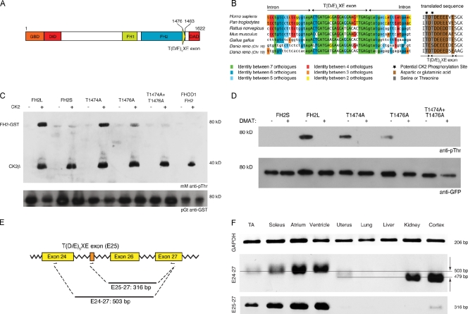
Figure 1.
FHOD3 contains a novel alternative exon at the end of its FH2 domain. (A) Domain layout of the human FHOD3 splice isoform expressing the T(D/E)5XE exon (exon 26 in humans and exon 25 in rodents; Fig. S1). GBD, GTPase-binding domain; DID, diaphanous inhibitory domain; FH1/FH2, formin homology domains 1 and 2. (B) The alternative T(D/E)5XE exon of FHOD3 encodes for an evolutionarily conserved stretch of eight mainly acidic amino acids that introduce CK2 consensus sites. Lowercase letters are intronic, and capital letters represent exonic sequences. Splice acceptor and donor sites are evolutionarily conserved in all seven orthologues, with the intronic bases AG on the 5′ end and GT on the 3′ end. (C) GST fusion proteins of the FHOD3-FH2 domain containing (FH2L) or excluding (FH2S) the alternative T(D/E)5XE exon, as well as threonine to alanine mutants of the predicted target threonines (T1474, T1476, or both), and the FHOD1-FH2 domain were tested for CK2 in vitro phosphorylation. FH2L, but not FH2S or FHOD1-FH2, was phosphorylated by CK2. Mutation of T1474 or T1476 to alanine reduced phosphorylation slightly or greatly, whereas mutation of both residues completely abolished the phosphorylation. mM, monoclonal mouse. (D) A similar phosphorylation pattern to that seen in vitro could also be demonstrated in COS-1 cells transfected with different GFP-tagged FHOD3 constructs and was abolished by treatment with 10 µM of the CK2 inhibitor DMAT for 24 h before lysis. The signal for GFP is shown as a loading control below. (E) Schematic view of the primer sets used for the RT-PCRs. (F) RT-PCR to test for tissue-specific expression of the alternative T(D/E)5XE exon. The amount of template mouse cDNAs was normalized against glycerine aldehyde 3-phosphate dehydrogenase (GAPDH). The specificity of the T(D/E)5XE exon for striated muscle can either be seen with primer set E25-27 or, because of the difference in the electrophoretic mobility, with primer set E24-27. The top line points out the 503-bp fragment, and the bottom line indicates the 479-bp fragment, which are also indicated by the arrows. TA, tibialis anterior.

