Figure 6.
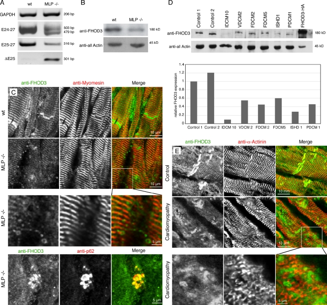
FHOD3 expression levels are reduced in the failing heart. (A) RT-PCR analysis of samples from MLP knockout hearts and wild type (wt) using primers for glycerine aldehyde 3-phosphate dehydrogenase (GAPDH) for standardization and primer sets for FHOD3, amplifying all splice variants (E24-27), amplifying variants specific for the T(D/E)5XE exon containing (E25-27), or amplifying exclusively the T(D/E)5XE exon excluding isoform (DE25). A dramatic change from splice isoforms containing the T(D/E)5XE exon to the isoform lacking this exon in hearts of MLP knockout mice can be detected. (B) The reduction of total FHOD3 (E24-27) mRNA in MLP hearts is mirrored by reduced FHOD3 protein levels in immunoblot analysis. (C and E) Immunofluorescence with an anti-FHOD3 antibody shows reduced staining in sections of MLP knockout (C) and human failing hearts (E), whereas the intensity of the antimyomesin and anti–α-actinin stainings are comparable. The reduction in muscle FHOD3 is accompanied by the appearance of autophagosome-like structures, which also colabel for p62 (C and E, inset magnifications; C, bottom stained with monoclonal mouse anti-p62). (D) Reduced expression of FHOD3 is also detected by immunoblot analysis in a range of human cardiomyopathies. A quantification of the blotting is shown below. Because of the scarcity of the material, the blotting was only repeated once. IDCM, idiopathic dilated cardiomyopathy; VDCM, ventricular dilated cardiomyopathy; FDCM, familial dilated cardiomyopathy; ISHD, ischemic heart disease; PDCM, perinatal dilated cardiomyopathy.

