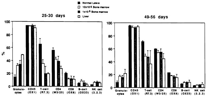
Figure 4.
Lineage of peripheral leukocytes shown in Figure 1. Granulocytes were determined on blood smear following α-naphthyl acetate esterase and hematoxylin staining. Other FACS analysis of percentage of subpopulations (mean ± SD) was by direct immunofluorescence with antibodies to surface markers, including OX1 (CD45), R7.3 (αβ TCR), W3/25 (CD4), OX8 (CD8), OX33 (B-cells), and NK3.2.3 (NK cells).

