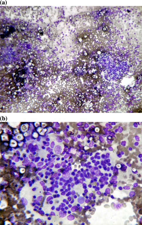
Fig. 3.
a, b Cytological smears 10× and 40× view stained with MGG stain showing cellular smears with predominant lymphocytic population. Lymphocytes are large and atypical with coarse chromatin and prominent nuclei, there is also presence of macrophages, occasional mature lymphocytes with RBCs in background. Cytological features suggestive of lymphoreticular malignancy suspicious of NHL (large cell type)

