Fig. 5.
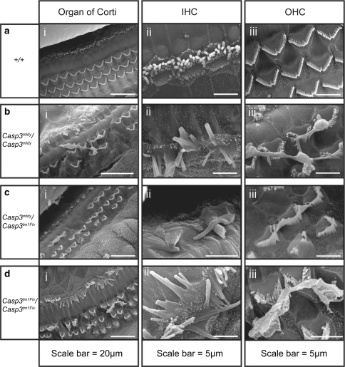
Scanning electron micrographs showing abnormal hair cell morphology in the cochlea of Casp3 mldy /Casp3 mldy (n = 3), Casp3 mldy /Casp3 tm1Flv (n = 2), Casp3 tm1Flv/Casp3 tm1Flv (n = 2), and +/+ mice (n = 2) aged 2–3 months. Panel (a i) shows a wild-type organ of Corti, and three well-organised rows of outer hair cells (OHC) and one row of inner hair cells (IHC) are visible. Casp3 mldy /Casp3 mldy (bi), Casp3 mldy / Casp3 tm1Flv (c i), and Casp3 tm1Flv/Casp3 tm1Flv (d i) display OHC degeneration and abnormalities of the stereocilia bundles of IHCs and OHCs. Panels (a ii)–(d ii) show higher-magnification images of the IHCs, and panels (a iii)–(d iii) show higher-magnification images of the OHCs. Casp3 mldy /+ (n = 3) and Casp3 tm1Flv/+ (n = 2) mice were also analysed (data not shown) but showed no differences to wild-type (see “Results”)

