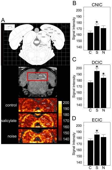
Figure 8. Effects of salicyate and noise exposure on MEMRI measured signal intensity in the inferior colliculus after three days of salicylate administration and 48 hrs post noise exposure.
Signal intensity was compared across groups (A–D). In the salicylate group, manganese accumulation was supernormal in the central nucleus of the inferior colliculus (B; CNIC; 174. 20±3.60; p = 0.03), as well as the dorsal cortex (C; DCIC; 195. 21±4.96; p = 0.005) and external cortex (D; ECIC; 186. 52±2.35; p = 0.02) when compared to controls. In the noise exposed group, only the DCIC had increased signal intensity (186. 35±2.17; p = 0.03). Asterisks denote significance, p≤0.05; error bars equal StDev, C – control, S – salicylate, N - noise exposed.

