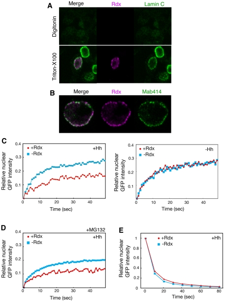
Figure 5. Rdx inhibited the nuclear import of Ci-155.
(A, B) Localization of Rdx at the nuclear pores. Clone-8 cells were transfected with the HA-Rdx expression vector, stained with anti-HA antibody (magenta), anti-lamin antibody (green) (A), or monoclonal antibody 414 (green) (B), after treatment with digitonin (upper in A) or Triton X-100 (lower in A and B), and were then analyzed by confocal microscopy. (C) Rdx inhibited the nuclear import of Ci-155 only in the presence of a strong Hh signal. FRAP experiments were performed using clone-8 cells transfected with the Ci-GFP and Hh expression vector, in the presence or absence of the Rdx expression plasmid (upper). The average of multiple experiments at each time point (n = 8 for –Rdx and n = 6 for +Rdx) is shown. Similar experiments were performed, with the exception of the absence of the Hh expression vector (lower). The average of multiple experiments at each time point (n = 8 for –Rdx and n = 10 for +Rdx) is shown. (D) Rdx inhibited the nuclear import of Ci-155 even in the presence of MG132. FRAP experiments were performed in the presence of the Hh expression palsmid as described above. Transfected cells were treated with MG132 (50 µM) for 5 h before harvesting the cells. (E) Rdx did not affect the nuclear export of Ci-155. FLIP experiments were performed using clone-8 cells transfected with the Ci-GFP and Hh expression vector, in the presence or absence of the Rdx expression plasmid. The decrease in nuclear Ci-GFP signal after cytosolic photobleaching was assessed, to monitor nuclear export. The average of multiple experiments at each time point (n = 6 for both –Rdx and +Rdx) is shown.

