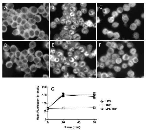
Figure 5.
TMP does not prevent nuclear translocation of NF-κB. Cells were either left untreated (A) or treated with LPS (1 μg/ml) for 20 (B) or 60 min (C) then stained. In panel D, cells were treated with 25 μM TMP for 60 min while panels D and F show treatments with LPS and TMP for 20 and 60 min, respectively. Following treatment cells were fixed, permeabilized and stained with anti-RelA Ab and a fluorescein coupled secondary Ab. Representative images from a single experiment are shown in A-F. For G, Photoshop (Adobe) was used to analyze images and determine mean fluorescence intensity for the nuclear region of 120 cells at each time point for each variable (20 cells from two fields from three independent experiments). Treatment with LPS and LPS in combination with TMP did not produce significant differences (p < 0.05, T-test).

