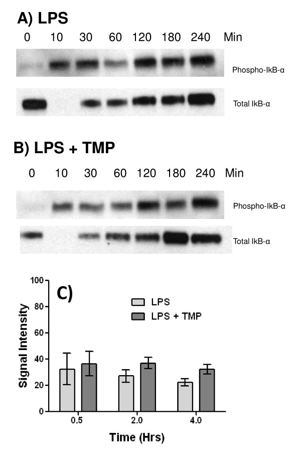
Figure 6.
TMP fails to affect IκB-α phosphorylation. RAW 264.7 cells were stimulated with either 1µg/ml of LPS (A) or 1µg/ml of LPS and 25µM TMP (B) for the indicated times. Lysates were prepared and analyzed by western blot with Abs specific for the phosphorylated serine 32 residue of IκB-α and total IκB-α. Representative experiments are shown in panels A and B. For densitometric analysis (C), phospho-IκB-α blots were scanned and band intensity determined using Photoshop. Values shown are means +/- SEM from three independent experiments. Treatment with LPS and LPS in combination with TMP did not produce significant differences (p < 0.05, T-test).

