Figure 8.
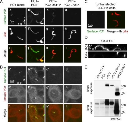
Coexpression with PC2 promotes PC1 localization to the apical and ciliary plasma membranes in polarized cells. LLC-PK cells stably expressing PC1 alone or together with PC2, PC2-D511V or PC2-L703X were grown to confluence and surface labeled with antibody directed against the N-terminal FLAG epitope (A, a–d and B, a′–d′). They were then permeabilized and labeled with an antibody directed against ciliary acetylated tubulin (A e-h), or the C-terminal HA epitope on PC1 (B, e′–h′). A small amount of PC1 was detectable on the primary cilium when PC1 was expressed alone (Aa). Expressing PC2 with PC1 dramatically increased the amount of PC1 at the ciliary membrane (Ab). Both PC2-D511V and PC2-L703X failed to stimulate PC1 delivery to the ciliary membrane (A, c and d). The anti-FLAG antibody gives no detectable ciliary background signal in untransfected cells (C). Images created by merging stacks of 20 successive Z plane confocal images reveal the extent to which PC1 is present at the apical and ciliary membranes (B, a′–d′) as well as the total amount of PC1 in the cells (B, e′–h′). Although all cell lines possess comparable amounts of total PC1 (B, e′–h′), PC1 is present at both the apical and ciliary membranes only when PC1 is expressed with PC2 (Bb′). A cross-section of the cells along the X-Z planes shows the presence of surface PC1 both at the apical membranes and in a pattern that colocalizes with that of the ciliary marker, an antibody directed against acetylated tubulin (E). A Western blot using an antibody directed against an N-terminal region of PC2 shows robust PC2 expression in the stable cell lines (E, top). A darker exposure shows low levels of endogenous PC2 in untransfected (WT) LLC-PK cells, and cells only transfected with PC1 (E, bottom).

