Figure 6.
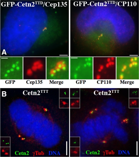
Centriole overduplication in GFP-Cetn2–expressing cells. (A) Extra Cetn2-containing foci contain several centriole markers. HeLa cells transfected GFP-Cetn2TTD and arrested in S-phase for 48 h were stained with antibodies against centriolar markers Cep135 and CP110. Shown are whole cell images (bar = 5 μm) and digitally magnified images of centrosomes (bar = 1 μm); green, GFP; red, Cep135 or CP110. (B) Extra Cetn2-containing foci recruit γ-Tubulin and participate in mitotic spindle assembly. HeLa cells were transfected with empty vector or untagged Cetn2 and analyzed with antibodies against Cetn2 and γ-Tubulin. Shown are cells with either pseudobipolar (left panel) or multipolar mitotic spindles from the Cetn2 transfection; green, Cetn2; red, γ-Tubulin. Bar = 5 μm. Insets show unmagnified images of spindle poles.

