
Figure 2. Detailed coupling pattern of dark-adapted OFF α-GCs.
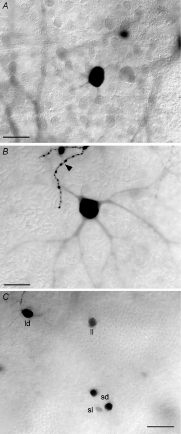
A, labelled soma of an OFF α-GC that is tracer coupled to a neighbouring OFF α-GC injected with Neurobiotin. Note that the dendritic arbour of the coupled cell is not labelled with only the primary dendrites partially visible. This was the typical labelling pattern for coupled OFF α-GCs in the dark-adapted rabbit retina. The soma injected α-GC is just out of view along the upper right margin of the panel. Scale bar = 25 μm. B, tracer coupled OFF α-GC that was particularly well labelled so that a sizeable portion of its dendritic arbour could be visualized. The arbour shows the radial branching pattern typical of α-GCs. The arrowhead indicates the well-labelled terminal dendritic branches of the OFF α-GC that was injected with Neurobiotin. Scale bar = 25 μm. C, the variability of the soma size and labelling intensity of the amacrine cells that are tracer coupled in OFF α-GCs. Based on these two parameters, we differentiated the coupled amacrine cells into four subtypes: large/dark (ld), large/light (ll), small/dark (sd), and small/light (sl). Scale bar = 25 μm.