
Figure 7. A series of confocal microscope images illustrating VGLUT1 terminals contacting a labelled interneuron and their relationship with GABA-containing terminals.
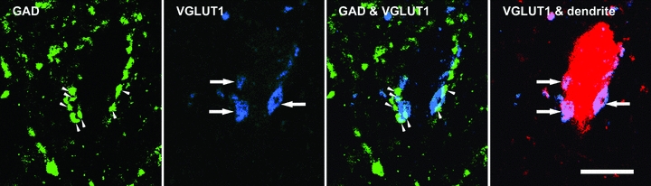
The left two panels show single optical sections through individual boutons (arrowheads and arrows). The dendrite of the postsynaptic interneuron is shown in red, immunoreactivity for GAD in green and VGLUT1 in blue. The right two panels are merged images confirming that the VGLUT1 terminals are associated with presynaptic GAD terminals and are in turn presynaptic to the labelled interneuronal dendrite. Scale bar = 10 μm.