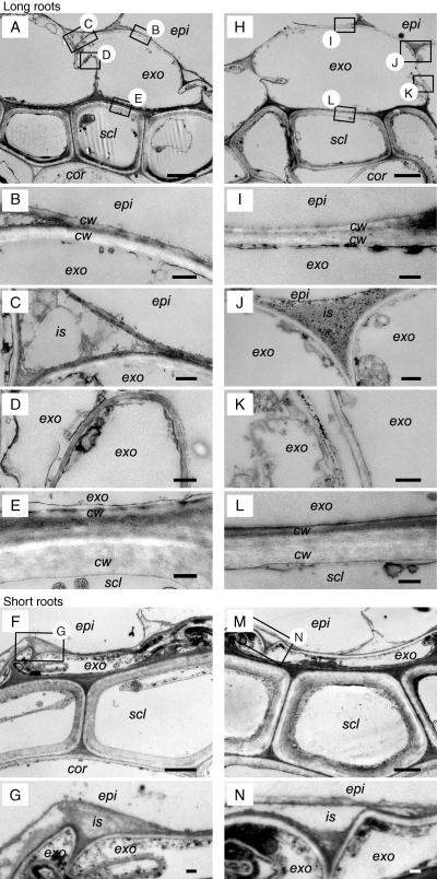
Fig. 3.
Comparison of the microstructure of the exodermis and the sclerenchyma in the basal part (15 mm below the root–shoot junction) of short or long adventitious roots grown continuously in aerated nutrient solution (short roots, F, G; long roots, A–E) or following transfer to stagnant deoxygenated agar nutrient solution for 48 h (short roots, M, N; long roots, H–L). Sections stained with uranyl acetate and lead citrate were observed by TEM. We observed the exodermis and sclerenchyma region (A, F, H, M; scale bar = 2 µm) and then specific cells at higher magnification (scale bar = 0·2 µm) for the epidermis side of the exodermis (B, C, G, I, J, N), between the exodermis cells (D, K), and also of the sclerenchyma side of the exodermis (E, L). Abbreviations: cor, cortex; cw, cell wall; epi, epidermis; exo, exodermis; is, intercellular space; scl, sclerenchyma. Plants were raised for 3–4 weeks in aerated nutrient solution, prior to transfer to stagnant deoxygenated agar nutrient solution for 48 h or aerated nutrient solution for 48 h. At the commencement of treatments the roots studied were short (65–90 mm) and long (105–130 mm) adventitious roots. Small boxes with letters in A and H and F and M indicate areas viewed at higher magnification and shown in the other panels of this figure.

