Figure 2.
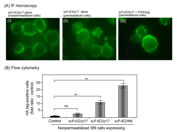
In situ analysis of scFvE2/p17 and scFvG2/p17 proteins in Sf9 cells. (A), Immunofluorescence (IF) microscopy. Sf9 cells expressing scFvE2/p17 alone (i, ii), or coexpressing scFvE2/p17 and Pr55Gag (iii), were harvested at 48 h pi and nonpermeabilized (i), or permeabilized with Triton X-100 (ii, iii). Cells were reacted with anti-HA tag monoclonal antibody followed by Alexa Fluor® 488-labeled complementary antibody. (B), Flow cytometry. Nonpermeabilized Sf9 cells expressing scFvE2/p17, scFvG2/p17 or the scFv-N18E2/M6 chimera were harvested at 48 h pi, reacted with antibodies as in (A), and analyzed by flow cytometry. Results shown were the proportion of HA tag-positive cells, expressed as the fold ratio over the values of control cells, attributed the value of 1. Control consisted of BVCAR-infected cells, i.e. cells expressing irrelevant membrane glycoprotein. BVCAR was a recombinant BV expressing the human CAR glycoprotein, and BVCAR-infected cells released CAR-displaying virions in the extracellular medium [41]. Average of three separate experiments, m ± SEM; (**), P < 0.01; ns, not significant.

