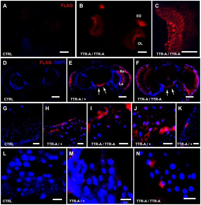
Figure 1. TTR localization pattern in transgenic flies.
Immunodetection of FLAG-TTR (red in A–N) with nuclear counterstaining (blue in D–N) on cryo-sections is shown. Third instar larval CNS with eye discs of control (A), and FLAGTTR-A/FLAGTTR-A (B, C) larvae show that the staining was specific for FLAG-TTR–expressing animals and restricted to the eye disk (ED) and Optic Lobe (OL) as expected for the GMR-Gal4 driver. A detail of FLAG-TTR expression in the terminals of retinal photoreceptors inside the OL of the brain is shown in C. Horizontal head sections of 14 days old control (D), FLAGTTR-A/+ (E) and FLAGTTR-A/FLAGTTR-A (F) adult flies showing FLAG-TTR localization in the retina (Re) and lamina (La) as well as in perineurial glia (arrows). The staining was absent in the head fat body of control flies (G) and present in FLAGTTR-A/+ (H, J), and FLAGTTR-A/FLAGTTR-A (I) flies. Detail of retina with TTR-A aggregates in FLAGTTR-A/+ flies (K). Thoracic fat body of control (L), and FLAGTTR-A/+ (M), and FLAGTTR-A/FLAGTTR-A flies (N). Aggregates of TTR-A were found in the retina and in fat body of head and thorax (Red “spots” in H–J and M–N). Scale bars, 100 µm in A–B, and D–F; 50 µm in C; and 20 µm in G–N. For a complete definition of the genotypes see Material and Methods.

